Atlas of Domestic Animal Neurological pathology and MRI
Goals and didactic concept
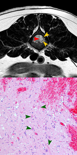
The present atlas was produced as an extension to the manual of "Veterinary Neuropathology, Essentials of Theory and Practice" (see below). The goal is to connect Diagnostic Imaging (MRI) and Neuropathology by comparing the gross and microscopic anatomy of neurological lesions. This approach should foster pattern recognition of neurological diseases, and should help to understand the morphologic background and nature of abnormal signal intensities in magnetic resonance images, the corresponding pathologies of abnormal signal hyper- and hypointensities, and of different contrast uptake patterns in different types of lesions.
This interactive learning program presents a series of neurological cases, including clinical findings, MRI with access to the original studies, gross and microscopic pathology. In a second step, the radiological report is presented, and typical features of the lesions are highlighted, in a third step direct correlation of MRI findings with pathology images is given.
Each case can be accessed in different ways: clinicians may first focus on neurologic signs by entering the atlas via "Cases", radiologists on the imaging studies by entering via "MRI", and pathologists on histopathological description and diagnosis by clicking on "Histology". Entering over "Image archive" allows search for specific diagnoses or disease group.
This atlas adresses
- Universities training veterinary specialists (residents) in the fields of neurology, radiology and pathology
- Veterinarians working in private clinics with special interest in neurology, MRI or pathology
- University students, doctoral students with special interest in these fields
Anatomy
- The anatomy of the brain can be reviewed on the following website:
http://vanat.cvm.umn.edu/mriBrainAtlas/
Contributers
- Johann Lang: Editor and Author diagnostic imaging; Division of Clinical Radiology, Department of Clinical Veterinary Sciences, Vetsuisse Faculty, University of Bern
- Daniela Gorgas: Author; Division of Clinical Radiology, Department of Clinical Veterinary Sciences, Vetsuisse Faculty, University of Bern
- Diana Henke: Author; Division of neurology, Department of Clinical Veterinary Sciences, Vetsuisse Faculty, University of Bern
- Anna Oevermann: Author neuropathology; Neurocenter, Department for clinical research, Vetsuisse Faculty, University of Bern
- Marc Vandevelde: Author neuropathology; Division of neurology, Department of Clinical Veterinary Sciences, Vetsuisse Faculty, University of Bern
- Aiko Matter: Coordination, programming and layout; Division of Clinical Radiology, Department of Clinical Veterinary Sciences, Vetsuisse Faculty, University of Bern
- Ulrich Woermann: Didactical concept, programming and layout; Unit of Instruction and Media AUM, Institute of Medical Education IML, University of Bern
- Dr. med. M. Rolli: Developer of content management system; Unit of Instruction and Media AUM, Institute of Medical Education IML, University of Bern
Acknowledgments:
- Our Diagnostic imaging technicians
- Our histology technicians
- Karine Gendron for proof reading the MRI reports
- Jeannine Brunner-Singh for support in developing the data base
Companion Website:
 This is a companion website of "Veterinary Neuropathology, Essentials of Theory and Practice" (Vandevelde, Higgins and Oevermann). At the end of each pathology report a reference to the corresponding pages of the book is included.